
Advances in Proteomics have Changed TPD Drug Discovery Pipelines
Proteome-wide measurements are changing how targeted protein degradation compounds move through drug discovery pipelines. Tandem mass tag (TMT) workflows remain widely used for multiplexed quantitative proteomics, providing deep coverage and an unbiased view of target degradation and proteome-wide selectivity. More recently, advances in data-independent acquisition (DIA), automated sample preparation, and high-speed instruments such as the Orbitrap Astral and timsTOF Ultra have dramatically increased throughput while maintaining deep proteome coverage [1].
Steger and colleagues recently demonstrated how these capabilities can be incorporated directly into molecular glue discovery, using a label-free DIA platform in 96-well format across two cell lines and using the data to evaluate structure-degradation relationships [2]. This example highlights how global proteomics has moved upstream in TPD pipelines, enabling increasingly large degrader and molecular glue libraries to be screened earlier in discovery, although the instrumentation and infrastructure required for high-throughput DIA remain a substantial investment.
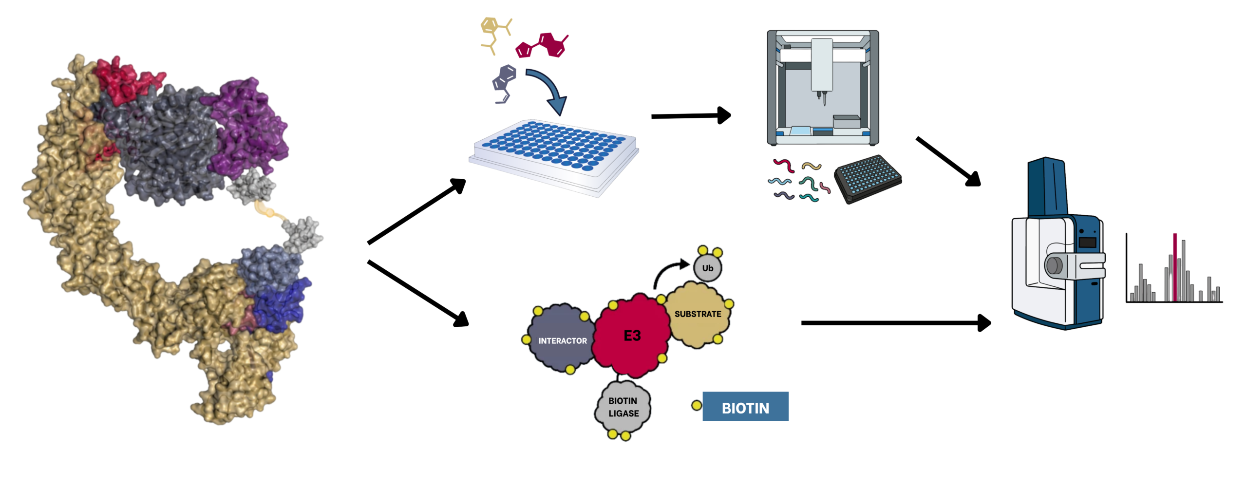
Proteomics has also shown value well beyond identifying proteins and measuring their relative abundance. Protein interaction and proximity proteomics provide complementary approaches for studying natural and chemically induced protein associations. BioID and related proximity-labeling methods use promiscuous biotin ligase mutants fused to proteins of interest to label nearby proteins with biotin. This allows for affinity enrichment and identification by mass spectrometry of stable and transient associations. These approaches were being applied to E3 ligase substrate discovery by 2015 and have since become useful tools for studying E3 interactomes and substrates [3,4]. Affinity-based approaches such as IP-MS instead identify proteins that co-purify with an E3 ligase and can be particularly useful for identifying compound-induced interactions. Yoon, Wachter and colleagues recently demonstrated the scalability of this strategy by simultaneously screening seven recombinant E3 ligases against pools of 320 compounds and cellular lysate, followed by deconvolution to identify the responsible E3 ligase and small molecule [5].
Identifying proximity or interaction does not necessarily establish that a protein is a substrate of the recruited E3. Recent methods including BioE3, E-STUB, Ub-POD, and the older UBAITs method attempt to bridge this gap by directly linking E3-specific ubiquitylation to proteomic identification [6-9]. Although their implementations differ, BioE3, E-STUB and Ub-POD each fuse wild-type BirA to an E3 ligase and coexpress it with ubiquitin tagged with a biotin acceptor peptide. Proximity between the E3 and engineered ubiquitin promotes selective biotinylation of ubiquitin that is transferred to substrates, allowing proteins ubiquitylated by the E3 to be enriched and identified by mass spectrometry. These methods provide different information compared to conventional proximity labeling – not simply proximity to an E3, but which proteins are ubiquitylated.
How much of this mechanistic information is needed during routine degrader discovery remains an important question. Global degradation proteomics provides the measurement closest to the desired pharmacology: the extent and selectivity of degradation. Proximity and E3-specific ubiquitylation proteomics provide additional layers of mechanistic information, but generally at lower throughput and with more specialized experimental requirements. Thus, these approaches may be most valuable downstream in a discovery cascade, where they can characterize promising compounds, distinguish productive from non-productive interactions, or help explain degrader action within the cellular context.
Proteomics can now generate increasingly detailed pictures of degrader activity in cells, but cellular measurements do not replace focused mechanistic experiments. Purified, reconstituted ubiquitylation systems provide an orthogonal approach for directly testing whether a proposed E3 ligase can ubiquitylate a target in the presence of a degrader. They can also validate the function of supplementary components, such as the priming E3 ligase ARIH1. As proteomic technologies continue to increase in both scale and scope, combining cellular discovery with biochemical validation provides a complementary path to comprehension.
Ternary complex (TC) formation does not guarantee productive ubiquitin transfer. If the target protein is degraded in cells, then the induced TC is likely capable of productive ubiquitylation, and optimization of the reconstituted system may be needed. On the other hand, if degradation is not observed in cells, alternative PROTAC exit vectors or linker characteristics may be needed to better position target lysines toward the ubiquitin-transfer machinery.
To address the former, start by evaluating the ubiquitylation machinery itself. E2 scouting can be particularly important, as different E2s can vary substantially in their ability to support initiation and chain extension with a given E3 system. Some E3-target pairs may require priming or cooperating ligases, such as ARIH1, while others require specific cofactors, post-translational modifications, or activation states for efficient activity.
Proteomics is the most direct starting point. Following treatment under conditions that promote target ubiquitylation, enrich the target protein and identify diGly-modified peptides by mass spectrometry. Proteasome inhibition can help preserve ubiquitylated species, and DUB inhibitors may be useful during cell lysis. If feasible, enriching the target before diGly peptide enrichment can improve sensitivity.
Candidate sites can then be validated orthogonally. Lysine mutants expressed in cells can be tested using quantitative degradation assays such as HiBiT or GFP-based measurements. Alternatively, recombinant mutant target proteins can be tested in a reconstituted ubiquitylation system to directly determine how individual sites or regions affect target ubiquitylation. Proteomics can therefore be particularly useful for narrowing the candidates before generating a large panel of recombinant mutants.
References
1. Lopes FBTP, Schlatzer D, Sudhadevi T, et al. What Does Next-Generation Mass Spectrometry Offer for Proteomics? A Comprehensive Platform Comparison. Journal of Proteome Research. 2026;25(4):1929-1940. doi:10.1021/acs.jproteome.5c01007.
2. Steger M, Nishiguchi G, Wu Q, et al. Unbiased mapping of cereblon neosubstrate landscape by high-throughput proteomics. Nature Communications. 2025;16:7773. doi:10.1038/s41467-025-62829-0.
3. Coyaud E, Mis M, Laurent EMN, et al. BioID-based Identification of Skp Cullin F-box (SCF)beta-TrCP1/2 E3 Ligase Substrates. Molecular & Cellular Proteomics. 2015;14(7):1781-1795. doi:10.1074/mcp.M114.045658.
4. Matsuhisa K, Sato S, Kaneko M. Identification of E3 Ubiquitin Ligase Substrates Using Biotin Ligase-Based Proximity Labeling Approaches. Biomedicines. 2025;13(4):854. doi:10.3390/biomedicines13040854.
5. Yoon H, Wachter F, Barrett KA, et al. DCAF11-dependent molecular glue degrader activated by glutathionylation. Nature. 2026. doi:10.1038/s41586-026-10873-1.
6. O’Connor HF, Lyon N, Leung JW, Agarwal P, et al. Ubiquitin‐Activated Interaction Traps (UBAITs) identify E3 ligase binding partners. EMBO Rep 16, 15 (2015). doi.org/10.15252/embr.201540620.
7. Barroso-Gomila O, Merino-Cacho L, Muratore V, et al. BioE3 identifies specific substrates of ubiquitin E3 ligases. Nature Communications. 2023;14:7656. doi:10.1038/s41467-023-43326-8.
8. Huang H-T, Lumpkin RJ, Tsai RW, et al. Ubiquitin-specific proximity labeling for the identification of E3 ligase substrates. Nature Chemical Biology. 2024;20(9):1227-1236. doi:10.1038/s41589-024-01590-9.
9. Mukhopadhyay U, Levantovsky S, Carusone TM, et al. A ubiquitin-specific, proximity-based labeling approach for the identification of ubiquitin ligase substrates. Science Advances. 2024;10(32):eadp3000. doi:10.1126/sciadv.adp3000.
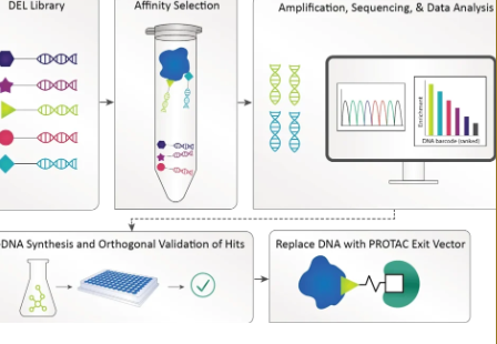
DNA-encoded libraries (DELs) are one example of a new discovery technology that is uniquely suited for degrader molecule discovery.
In Vitro Proximity and Ubiquitylation Assays for CRBN, VHL and Emerging Ligases of Interest in TPD Introduction Trilogy offers top-quality products and custom services. Our innovative in vitro assays complement cell-based methods, providing valuable insights in TPD workflows.
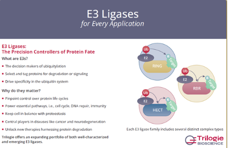
Well-characterized and emerging E3 ligase reagents to support ubiquitin biology, targeted protein degradation, mechanistic enzymology, and drug discovery research.